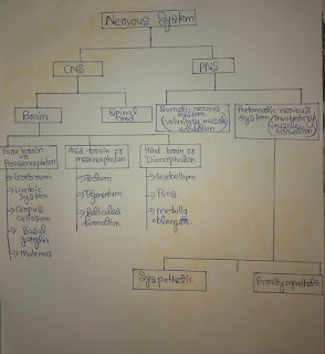
What is nervous system ? What is CNS ? What is PNS ? Structure or brain , spinal cord , ANS ? SNS?
Nervous system :-
The nervous system is divided into two parts which is :- a central nervous system [CNS] and peripheral nervous system [PNS]. The central nervous system is consists of brain and spinal cord, which lies within the bony cases of the skull and spine. The part of the nervous system outside the skull and spine make up the peripheral nervous system [PNS].
Central nervous system [CNS] :- It is the part of nervous system. It controls most functions of the body and mind. It consists of two parts : the brain and spinal cord. It is referred to as "central" because it combines information from the entire body and coordinates activity across the whole organism. It receives sensory information from the nervous system and control the body's responses.
Brain :- Human brain is the center of human nervous system, enclosed in the cranium. It is one of the largest and most complex organ in the human body. It receives signals from the body's sensory organs and outputs information to the muscles.
Structure of brain:- Our body is only 2% of our body weight, it consumes about 25% of our body's oxygen and 70% of it's glucose. Brain never rest's , it's rate of energy metabolism is relatively constant day and night. In fact, in our dreams the brain's metabolic rate actually increase slightly [ Hobson,1996]
A. Fore Brain [ prosencephalon] :- The most rostral of the 3 major division of the brain, includes the Telencephalon and diencephalon.
- Integration of olfactory information with others sense
- Regulation of body temperature, reproduction, eating, emotion
- Learning and memory in mammals
- Cerebral cortex consists mostly of glia in the cell bodies, dendrites and interconnecting axons of neurons. Because cell predominate, the cerebral cortex has a grayish brown appearance and it is called grey matter.
- White matter the large concentration of myelin around the axon give the tissue an opaque white appearance hence the term called white matter
- In human the cerebral cortex is gently furrowed. These convolution, consisting of sulci, fissures and gyri.
- Sulcus [ plural : sulci ] :- A groove in the surface of cerebral hemisphere, smaller than a fissure.
- Fissure :- A major groove in the surface of brain, larger than a sulcus. It separates each cerebral hemisphere into 4 lobes [ frontal, parietal, temporal, occipital ]. There are four fissures which are as follows :-
- Longitudinal fissure :- The 2 cerebral hemispheres are almost completely separated by the largest of fissures.
- Central fissure :- It is a prominent landmark of the brain separating the parietal lobe from the frontal lobe and the primary motor cortex from the primary somatosensory cortex.
- Lateral fissure [ sylvian fissure ] :- A deep fissure in each hemisphere that separates the frontal and parietal lobe from temporal lobe. The insular cortex or insula lies deep within the lateral fissure.
- Parieto-occipital fissure:- It separates the parietal and occipital lobes.
- Precentral gyri :- Found on the lateral surface of the frontal lobe and acts as the primary motor cortex of the brain.
- Postcentral gyri :- It contains somatic sensory cortex.
- Superior temporal gyri :- It contains auditory cortex. In fact 2-3rd of the surface of cortex is hidden in groove.
- Primary motor cortex :- It controls 600 or more muscles involved involuntary body movements. It lies at the back of the frontal lobe adjacent to the central fissure. Because the nerve tract from the primary motor cortex cross over at the level of medulla, each hemisphere governs movements on the opposite side of the body. Thus serve damage to the right motor cortex would produce paralysis in the left side of the body and vise versa. If we electrically stimulate a particular point on the motor cortex, movement occurs in the muscle governed by the part of the cortex.
- Primary somatosensory cortex:- With the exception of taste and smell, it receives input from our sensory receptor and gives rise to our sensation of heat, touch, cold and to our sense balance and body movement. It lies at the front portion of the parietal lobe just behind the primary motor cortex, separated from it by the central fissure. Each side of the body sends sensory input to the opposite hemisphere.
- Primary visual cortex:- It receives visual formation located at the back of the brain, on the inner surface of the cerebral hemisphere- primarily on the upper and lower banks of the calcarine fissure.
- Primary auditory cortex:- It receives auditory information located below surface of lateral fissure in the side of brain.
- Sensory association cortex:- Each primary sensory area of the cerebral cortex send information to adjacent regions called sensory association cortex. It is of four types:-
- Auditory association cortex
- Visual association cortex
- Somatosensory association cortex
- Motor association cortex
- The frontal lobe :- It includes everything in front of the central sulcus. It is the part of the brain that control important cognition skills in humans, such as emotional expression, problem solving, memory, language, judgement, and sexual behavior. It is in essence, the "central panel" of our personality and our ability to communicate. Complex chain of motor movement also seem to be controlled by the frontal lobes. MRI studies have shown that the frontal area is the most common region of injury following mild to moderate brain injury. There are important asymmetrical difference in the frontal lobes. The left frontal lobes is involved in controlling language related abilities, whereas the right frontal lobe plays a role in non-verbal abilities.
- Loss of simple movement of various body parts [paralysis]
- Inability to plan a sequence of complex movement needed to complete multi-shaped tests, such as making coffee[sequencing]
- Loss of spontaneity in interacting with other's
- Inability to express language
- The parietal lobe:- It is posterior to the central sulcus and frontal lobes, superior to occipital lobes and above the temporal lobes. The parietal lobes are involved in a number of important functions in the body. One of the main functions is to receive and process sensory information from all over the body. Neuron in the parietal lobes receive touch, visual and other sensory information from a part of the brain called the thalamus. The parietal lobes process the information and help us to identify object by touch.
- The first function integrates sensory information to form a single perception [ cognition ]
- The second function construct a spatial coordinate system to represent the world around us.
- Distinguishing between 2 points, even without visual input
- Localizing touch: When we touch any object with any part of our body, our parietal lobe enables us to feel the sensation at the site of the touch
- Integration sensory information from most region of the body
- Visuospatial navigation and reasoning: When we read a map, follow direction or prevent ourselves from tripping over a unexpected obstacles, our parietal lobe is involved. The parietal lobe is also vital for proprioception.
- Some visual function, in conjunction with the occipital lobe
- Assessing numerical relationship, including the number of object we see
- Assessing size, shape and orientation in space of both visible stimuli and objects we remember encountering
- Parietal lobe, Right- Damage to this area can cause visuospatial deficits [ e.g., the patient may have difficulty in finding their way around new or even familiar place ]. We are unable to care our body
- Parietal lobe, Left- Damage to this area may disrupt a person's ability to understand spoken or written language
- Damage that crosses both parietal lobe, leads to a condition called Balint's syndrome, which impedes motor skills and visual attention. People with Balint's syndrome may not be able to volunteer the eye.
- The Temporal lobe :- It is located posterior to the frontal lobe and inferior to the parietal lobe and sylvian fissure. The temporal lobe of the brain is often referred to as the neocortex. The temporal lobe is 2nd largest lobe[the frontal lobe is the largest].
- Left temporal lobe:- involved in understanding language and learning and remembering verbal information. It is dominate lobe.
- Right temporal lobe:- The non-dominant lobe, involve in learning and remembering non-verbal information.
- Auditory processing: one of the major function to receive any sound process those signal and tell us what they mean
- Speech/language recognition: A complex within the temporal lobe called auditory complex is responsible for helping us hear speech, understand what's being said.
- Speech generation: The temporal lobe and other parts of the brain work together to help us process and understand visual information and the temporal lobe also help us actually speak the words we what to say
- Memory: The other major function of temporal lobe is memory, specifically auditory, olfactory and visual memories
- Visual understanding: while the occipital lobe and other parts of the brain allow temporal lobe that help us remember and name things
- Hippocampus:- 2 hippocampi are found in each temporal lobe below the cerebral cortex. Involved in forming and retrieving memories. Damage in it can result in the sever memory impairment for recent events. The importance of hippocampus in this respect was solidified in 20th century by the case of a patient named Henry Malaison. He under went surgery to treat severe epilepsy in his late twenties. In that surgery, much of his hippocampi were either removed or damage. The surgery was successful in controlling Malaison's seizures, But afterward the suffered from sever anterograde amnesia.
- Amygdala:- 2 Amygdala are located close to the hippocampus, in the frontal portion of the temporal lobe of each side. It helps us to organize motivational and emotional response patters, particularly those linked to aggression and fear. Electrically stimulating certain area of the amygdala causes animals to evoke and immediate aggressive response. Stimulating of other area results in a fearful inability to respond aggressively, even in self defence .
- Corpus callosum:- It is a neural bridge consisting of white myelinated fibers that act as major communication link between the 2 hemisphere and allows them to function as a single units. The corpus callosum is the largest fiber bundle in the brain, containing nearly 200 million axons.
- Transmission of visual information
- Transmission of somatosensory information
- To integrate information
- Two hemisphere communicate with one another through the corpus callosum
- If it cut or damage produce 2 different and largely independent minds in the same person. It is split brain condition.
- From interior to posterior the corpus callosum can be divide into region known as rostrum, genus body and splenium.
- Occipital lobe :- The occipital lobe is important to being able correctly understand what your eyes are seeing. These lobes have to be very fast to press the rapid information that our eyes are sending.
- Mapping the visual world, which helps with both spatial reasoning and visual memory, most vision involves some type of memory
- Assessing distance, size, and depth
- Deferming color properties of the item in the visual field
- Transmitting visual information to other brain region
- It is located above the mid brain
- Resemble like 2 small football, one in each cerebral hemisphere
- It has sometimes been linked to a switch board that organize input from sensory organs and ruts them to the appropriate area of the brain.
- The visual, auditory and body sense[ balance and equilibrium] all have major relay stations in the thalamus
- Individual with disrupted functioning the thalamus often experience a higher confusing world.
- Tectum:- It is located in dorsal portion of mid brain, and divided into 2 parts:-
- Superior colliculi: part of visual system
- Inferior colliculi : part of auditory system
- It act as a sentry altering higher centers of the brain the messages are coming and then either blocking those messages or allowing them to go forward
- Consciousness, sleep, wakefulness, and attention
- Researches discovered that electrical stimulation of different portions of the reticular formation can produce instant sleep in a wakeful cat and sudden wakefulness in a sleeping animal
- Ascending part :- Sends input to higher regions of the brain to alert it. Without reticular stimulation of higher brain regions, sensory messages do not register in conscious awareness even though the nerve impulses may reach the appropriate higher are of brain. It is as if the brain is not awake enough to notice them.
- Descending part :- It serve as a kind of gate through which some inputs are admitted while others are blocked out by signals coming down from higher brain centers. [ Van Zomeren and Brouwer 1994]. Sever damage to reticular formation can produce a permanent coma.
- It plays role in heart rates, respiration and skeletal muscle tonus.
- The medulla is also a 2 way through fare. for all the sensory and motor nerve tracts coming up from the spinal chord and descending from the brain. Most of there tract cross over within the medulla.
- It concerned with muscular movement coordination, but it also plays a role in learning and memory.
- Specific motor movement are initiated in higher brain centers, but their timing and coordination depend on cerebellum. [ Tatch et al., 1992 ]
- The input function that enable us to sense what is going on inside and outside our bodies.
- The output that enable us to respond with our muscles and glands.
- Somatic nervous system :- SNS contain of sensory neurons that are specialized to transmit message from the eyes, ear and other sensory and motor neurons that send messages from the brain and spinal cord to the muscles that control our voluntary movement.
- The axon of sensory neurons group together and make sensory nerves
- The axon of motor neuron group together and make motor nerves
- Inside the brain and spinal cord, nerves are called tracts.
- Sympathetic nervous system :- It has an activation arousal function and it tends to act as a total unit.


Good knowledge
ReplyDeleteThank you mam
ReplyDeleteBhot achha content he
Aapki post ka bhot dino se wait krra tha so helpful mam
ReplyDeleteVery helpful
ReplyDeleteThank you mam
ReplyDeleteHelpful content 👍👍
ReplyDelete